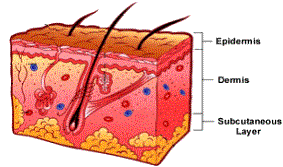

The anatomy of the skin around the eyes, also referred to as the adnexa is unique to the face and body. In order to properly care for the skin around the eyes, it is important to understand not only the anatomy of this area, but also the process of skin cell renewal.
| Eyelid skin is composed of several layers. The deepest, the subcutaneous layer contains a thin layer of fascia which lies on top of the orbicularis muscle, a muscle that allows the eyelid to move. Next, the dermis, which forms the support layer of the skin, is made up of threadlike proteins including bundles of elastin and collagen, fibroblasts, nerves and vessels. The top layer, the epidermis, is made up of basal cells, melanocytes, Langerhans cells, keratinocytes and on top, the dead cell layer (also known as the stratum corneum) made up of corneocytes. The epidermal layer gives the skin its appearance, color, suppleness, texture, and health. |  |
| Basal cells reproduce new cells every few days. As these cells migrate upward, they become drier and flatter. Once they reach the surface of the skin, they are no longer alive, and are referred to as corneocytes. This process of migration from basal cell to corneocytes is what gives the epidermis the ability to regenerate itself. This skin renewal process is known as desquamation. Desquamation is an ongoing process that takes about two seeks in a young person, and significantly longer – about 37 days for individuals over 50. The build up of corneocytes gives skin a callous or dry, aged and thickened look. The skin feels and looks rough and its ability to retain water becomes impaired. |
Appointment
Request
Please call for an appointment: 972-869-6900,
use our book now online service or email.
Our Favorite
Links
Please "like" us on Facebook!
fb.com/Skin.Technology.SherryModern portrait photography with an artistic flair.
pattyfoppen.com